Liver CT Image Segmentation
By training the AI model of medical image segmentation, the liver tissue
image is segmented from the original medical image to reduce the burden of clinicians manually
sketching the target area of medical image.
Background
- With the rapid development and popularization of medical imaging equipment, imaging technology has been widely used in clinical practice and has become an indispensable auxiliary means for disease diagnosis, surgical planning, prognosis evaluation and follow-up. Medical images often play a vital role in the process of diagnosis and treatment. Therefore, medical images have become one of the most important sources of evidence for clinical analysis and medical intervention. Medical image segmentation can extract key information from specific tissue images, which is a key step to realize medical image visualization. The segmented image is provided to doctors for quantitative analysis of tissue volume, diagnosis, localization of pathological changes, description of anatomical structure, treatment plan and other different tasks.
Prepare Dataset
- First step: Specify the application scenario. In this example, the focus areas of different organs in the image are divided.
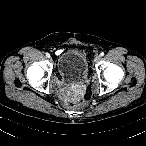
- Second step: It is recommended to prepare at least 200 medical images. For example:

PS: Click here to download a sample dataset.
If you need a complete dataset, please send mail to hpchelp@lenovo.com
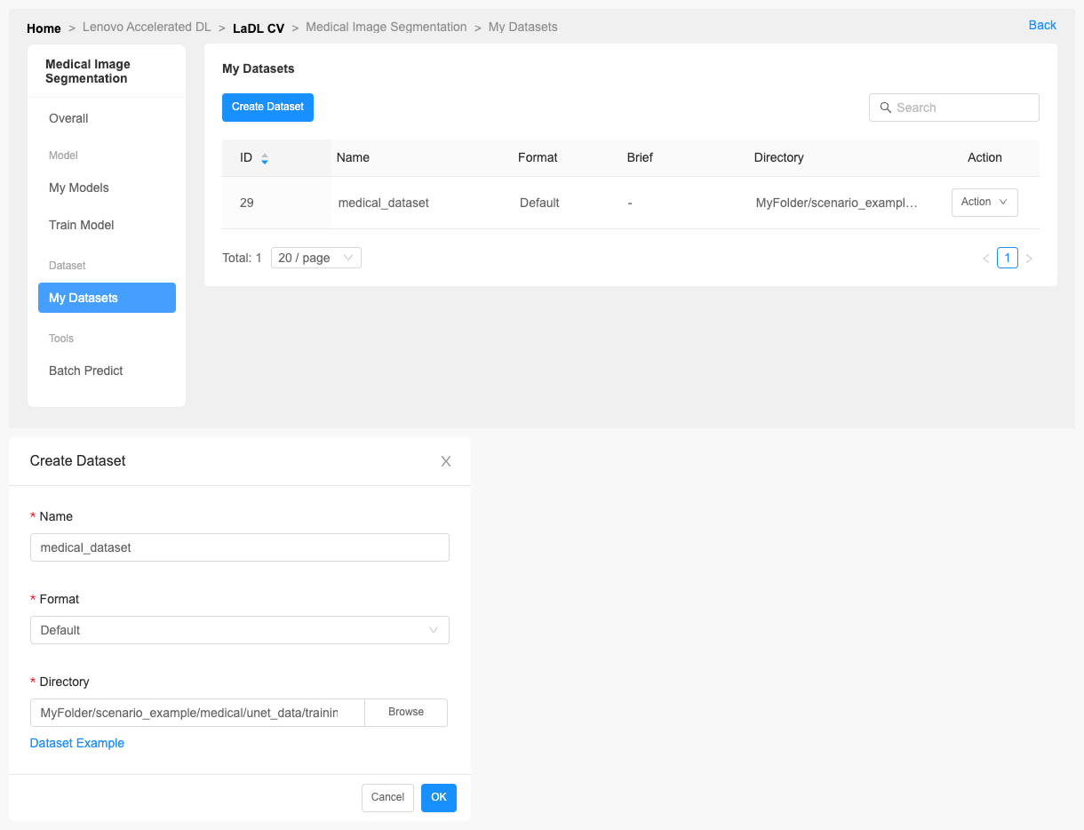
- Third step: Create a dataset through LiCO.
1.Click [Create Dataset].
Train Model
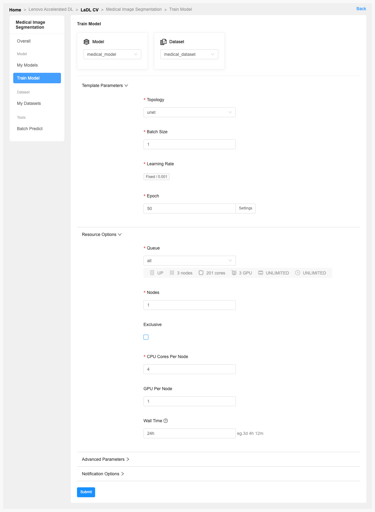
- When your dataset preparation is complete, you can click [Create Model] to complete the model creation.
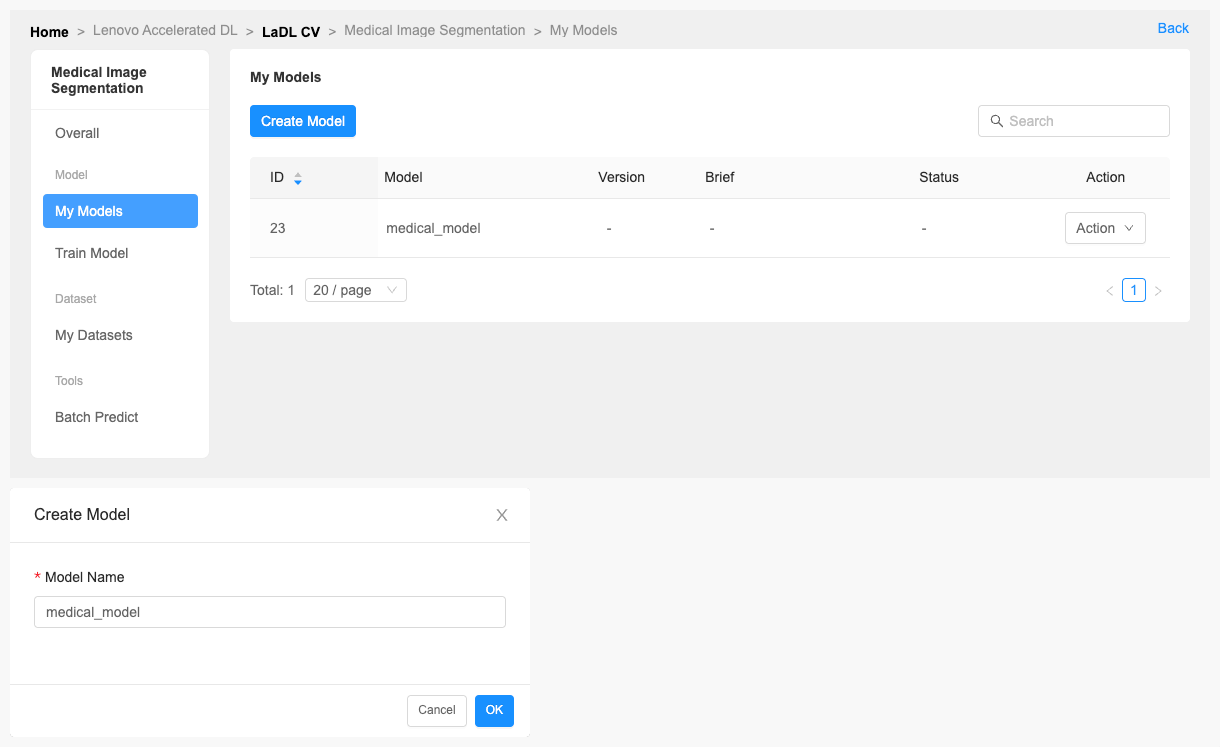
- And then click [Train Model] to start training.
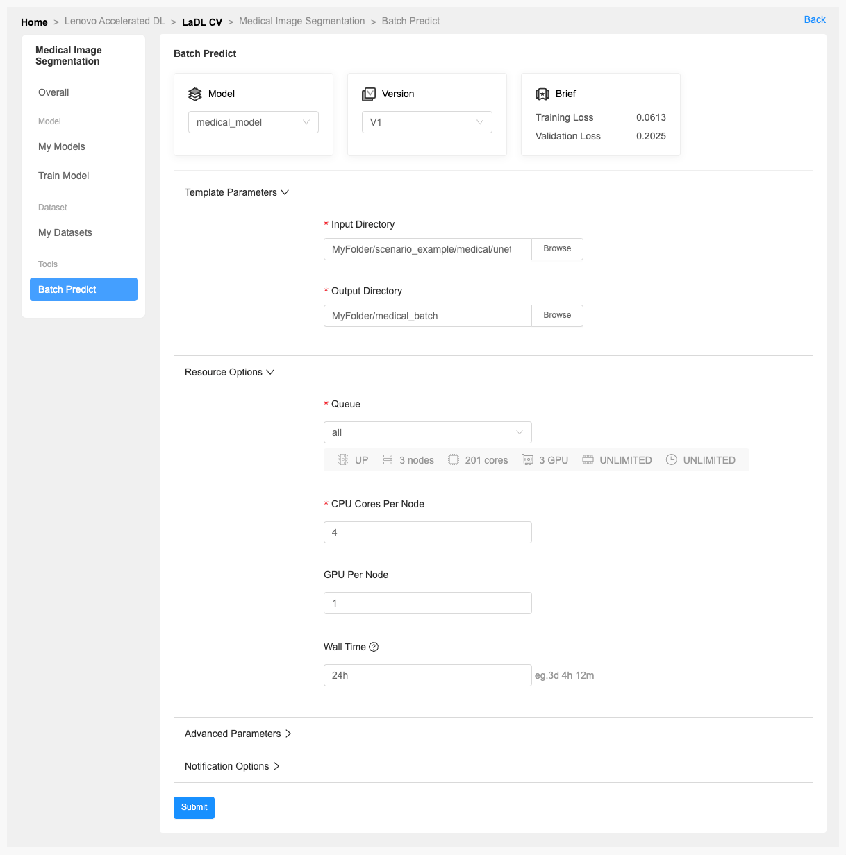
- You can also predict multiple images by [Batch Predict].
